Restless Legs Syndrome

Restless Legs Syndrome (RLS), also known as Willis-Ekbom Disease, is a neurological disorder that manifests as an irresistible urge to move the legs, often accompanied by uncomfortable sensations. The condition affects individuals of all ages and can significantly impact their quality of life. Despite its prevalence, the exact cause of RLS remains elusive, making it a challenging disorder to understand and manage effectively.

Symptoms:

The hallmark symptom of RLS is the compelling urge to move the legs, typically triggered by rest or inactivity. Individuals with RLS often describe uncomfortable sensations such as tingling, crawling, or itching deep within their legs. These sensations intensify during the evening and night, leading to difficulty falling or staying asleep. The discomfort associated with RLS can vary widely, ranging from mild to severe, and may significantly disrupt daily activities.

Prevalence:

RLS is a common disorder, with estimates suggesting that up to 10% of the population may be affected. The prevalence increases with age, and women are more commonly affected than men. While RLS can occur at any age, it often goes undiagnosed, as individuals may not recognize the symptoms or may be hesitant to discuss them with healthcare professionals.

Etiology:

The precise cause of RLS remains unclear, but a combination of genetic, environmental, and neurological factors is believed to contribute to its development. Research suggests a strong familial predisposition, indicating a genetic component. Additionally, abnormalities in dopamine, a neurotransmitter involved in movement control, have been implicated in the pathophysiology of RLS. Iron deficiency, pregnancy, and certain medications are recognized as secondary triggers or exacerbating factors.

Diagnosis:

Diagnosing RLS can be challenging due to its subjective nature and the absence of definitive laboratory tests. Healthcare professionals typically rely on a thorough clinical history and evaluation of symptoms. The International Restless Legs Syndrome Study Group has established criteria for diagnosing RLS, emphasizing the characteristic urge to move the legs and the relief experienced with movement.

Management:

While there is no cure for RLS, various treatment options aim to alleviate symptoms and improve quality of life. Non-pharmacological approaches include lifestyle modifications, such as regular exercise and avoidance of stimulants like caffeine and nicotine. Medications that affect dopamine levels in the brain, such as dopaminergic agents, are commonly prescribed. Iron supplementation may be recommended for individuals with identified deficiencies.

Conclusion:

Restless Legs Syndrome remains a complex and enigmatic disorder that poses challenges for both patients and healthcare professionals. Advances in research continue to shed light on its underlying mechanisms, offering hope for more targeted and effective treatments in the future. Increased awareness, improved diagnostic criteria, and a multidisciplinary approach are essential in addressing the impact of RLS on individuals’ lives and advancing our understanding of this intriguing neurological disorder.

The Foot Function Index

The Foot Function Index (FFI) is a self-report questionnaire or assessment tool used to evaluate the functional status and impact of foot-related conditions on a person’s daily life. It helps healthcare professionals and researchers assess the severity of foot problems, track changes over time, and monitor the effectiveness of treatments or interventions.

The FFI consists of three subscales or sections:

  1. Foot Pain (FFI-Pain): This section assesses the severity and impact of foot pain on a person’s daily activities. It includes questions related to the location, intensity, and frequency of foot pain, as well as how pain affects activities like walking, standing, and participating in sports or recreational activities.
  2. Foot Function (FFI-Function): This section focuses on the functional limitations caused by foot problems. It includes questions about a person’s ability to perform various activities, such as walking, climbing stairs, and participating in social and recreational activities, while considering the presence of foot issues.
  3. Foot-Related Health (FFI-Health): This section evaluates the impact of foot problems on overall health and well-being. It may include questions about emotional and psychological aspects related to foot conditions, as well as overall satisfaction with foot health.

The FFI is usually administered in the form of a self-administered questionnaire, and patients are asked to rate their responses on a scale (e.g., 0 to 100), with higher scores indicating greater functional limitations and more severe symptoms. Healthcare professionals can use the FFI to assess the impact of foot conditions on a patient’s quality of life and to guide treatment decisions. It is particularly useful in clinical settings for conditions such as plantar fasciitis, bunions, arthritis, and other foot-related issues

Rheumatoid arthritis and the Foot

Rheumatoid arthritis (RA) is a chronic autoimmune disease that primarily affects the joints, causing inflammation and pain. It can affect any joint in the body, including those in the feet. When RA affects the foot, it can lead to various symptoms and complications.

  1. Joint Inflammation: RA typically starts with inflammation in the synovium, the lining of the joint, which can lead to joint pain, swelling, and stiffness. In the feet, commonly affected joints include the smaller joints, such as those in the toes and the midfoot.
  2. Deformities: Over time, untreated or poorly managed RA can lead to joint deformities in the feet. Common deformities include hammer toes (toes that become permanently bent), claw toes (toes that curl downward), and bunions (a bony bump at the base of the big toe).
  3. Joint Erosion: RA can cause the cartilage and bone within the affected joints to erode, leading to a loss of joint space. This can result in reduced joint mobility and pain.
  4. Tendon Involvement: RA can also affect the tendons in the foot, leading to inflammation and weakening. Tendon involvement can result in problems like Achilles tendonitis or posterior tibial tendon dysfunction.
  5. Nodules: Some individuals with RA may develop subcutaneous nodules, which are firm lumps that can form on the bottom of the foot or elsewhere. These nodules are a sign of more severe RA and can be painful.
  6. Pain and Discomfort: Foot pain and discomfort are common symptoms of RA in the feet. This pain can make it difficult to walk or wear certain types of footwear.
  7. Limited Mobility: As the disease progresses and the joints are affected, it can limit your mobility and ability to perform daily activities. Walking and standing for extended periods can become challenging.
  8. Ulcers and Skin Problems: Reduced circulation and sensation in the feet, combined with deformities, can increase the risk of foot ulcers and skin problems. It’s important to care for your feet and inspect them regularly for any signs of issues.
  9. Impact on Gait: RA can affect your gait and the way you walk due to joint pain and deformities. This altered gait can, in turn, lead to other musculoskeletal problems and discomfort in other parts of the body.
  10. Treatment: Managing RA in the foot involves a combination of medication, physical therapy, and lifestyle changes. Medications may include disease-modifying antirheumatic drugs (DMARDs) to slow the progression of the disease, as well as pain relievers and anti-inflammatory drugs. Physical therapy can help maintain joint function and mobility. Custom orthotics or supportive footwear may be recommended to alleviate foot pain and improve comfort.

Early diagnosis and treatment are crucial to manage RA effectively and minimize its impact on the feet and overall quality of life. If you suspect you have rheumatoid arthritis or are experiencing foot pain and other symptoms, consult a healthcare professional for an accurate diagnosis and treatment plan.

Osteoarthritis and the Foot

Osteoarthritis is a common degenerative joint disease that affects millions of individuals worldwide. While commonly associated with aging, it can also strike younger individuals due to various factors, including genetics, injury, or repetitive stress. One area of the body that frequently bears the brunt of osteoarthritis is the foot. The complex structure of the foot and its continuous weight-bearing function make it susceptible to the development of osteoarthritis. In this essay, we will explore the intricacies of osteoarthritis, its impact on the foot, and the various treatment options available.

Understanding Osteoarthritis

Osteoarthritis, often referred to as “wear and tear” arthritis, is a chronic joint disorder characterized by the progressive degradation of the articular cartilage that covers the ends of bones. This condition can affect any joint in the body, but the foot is a common site for osteoarthritis due to its role in supporting the entire body’s weight and facilitating locomotion. In the foot, the weight-bearing joints most susceptible to osteoarthritis include the ankle, subtalar joint, and the metatarsophalangeal (MTP) joints (hallux rigidus).

The pathogenesis of osteoarthritis involves a complex interplay of genetic and environmental factors. While age is a primary risk factor, it is not the sole determinant. Genetics plays a role in a person’s susceptibility to the disease, and injury or trauma can accelerate its development. In the foot, osteoarthritis often develops gradually, with individuals experiencing symptoms such as pain, stiffness, and decreased range of motion.

Impact of Osteoarthritis on the Foot

  1. Pain and Stiffness: Osteoarthritis in the foot typically manifests as pain, often localized in the affected joints. This pain may worsen with activity and improve with rest. Stiffness in the foot, particularly in the morning or after periods of inactivity, is a common complaint.
  2. Decreased Range of Motion: As osteoarthritis progresses, individuals may notice a reduction in the range of motion in their foot joints. This can impact their ability to walk, stand, and perform daily activities.
  3. Swelling: Swelling, often referred to as effusion, can occur in the affected joints. This can be accompanied by warmth and tenderness, making it uncomfortable for individuals to put weight on their foot.
  4. Deformities: In severe cases of foot osteoarthritis, joint deformities may develop. This can lead to the misalignment of bones and the formation of bony growths known as osteophytes, which can further exacerbate pain and reduce function.
  5. Altered Gait: Pain and stiffness associated with foot osteoarthritis can alter an individual’s gait. This compensation for pain can lead to other musculoskeletal issues, such as knee or hip problems.

Diagnosis of Foot Osteoarthritis

Diagnosing foot osteoarthritis involves a thorough clinical examination and, in some cases, medical imaging. The healthcare provider will assess the patient’s medical history, symptoms, and perform a physical examination to identify the affected joints and evaluate their range of motion. Medical imaging techniques, such as X-rays, may be employed to confirm the diagnosis and assess the severity of osteoarthritis by examining joint space narrowing, osteophyte formation, and joint alignment.

Treatment Options

  1. Conservative Management:
    a. Pain Management: Non-steroidal anti-inflammatory drugs (NSAIDs) and analgesics can provide relief from pain and inflammation.
    b. Physical Therapy: Physical therapy can help improve joint flexibility, strengthen surrounding muscles, and promote a better gait.
    c. Foot Orthotics: Custom orthotic devices can help distribute pressure more evenly across the foot, reducing the strain on affected joints.
    d. Shoe stiffening inserts
    e. Weight Management: Maintaining a healthy weight can alleviate stress on the foot, reducing the progression of osteoarthritis.
  2. Assistive Devices:
    a. Bracing: Ankle braces or foot orthoses can provide support and reduce the risk of further joint damage.
    b. Canes or Crutches: These assistive devices can help reduce the load on the foot during walking.
  3. Injections:
    a. Corticosteroid Injections: Intra-articular corticosteroid injections can offer temporary pain relief by reducing inflammation in the affected joints.
    b. Hyaluronic Acid Injections: These injections can provide lubrication and cushioning for the joint, potentially alleviating pain.
  4. Surgical Interventions:
    a. Arthroscopy: Minimally invasive arthroscopic procedures can be used to clean out debris and osteophytes from the affected joints.
    b. Joint Fusion: In cases of severe osteoarthritis, joint fusion can be performed to permanently stabilize and relieve pain in the joint.
    c. Joint Replacement: Total joint replacement surgery, such as ankle arthroplasty, may be recommended for end-stage osteoarthritis to replace the damaged joint with an artificial implant.

Prevention and Lifestyle Modifications

While osteoarthritis is not always preventable, certain lifestyle modifications can reduce the risk and slow the progression of the condition. Maintaining a healthy weight, wearing appropriate footwear, and engaging in regular low-impact exercise can help support the foot’s overall health. It’s also essential to protect the feet from injuries and trauma, as these can increase the likelihood of osteoarthritis development.

Osteoarthritis in the foot is a significant challenge, affecting an individual’s mobility and overall quality of life. While there is no cure for this degenerative joint disease, effective management strategies are available to alleviate symptoms and improve function. Early diagnosis, along with a comprehensive treatment plan that may include conservative measures, assistive devices, injections, or surgical interventions, can make a substantial difference in the lives of those suffering from foot osteoarthritis. Furthermore, adopting a healthy lifestyle and taking preventive measures can help reduce the risk of developing this condition and its impact on the foot. As research continues, advances in the understanding and management of osteoarthritis in the foot will provide hope for a better future for affected individuals.

Gout

Gout is among those historic conditions as there are quite a few mentions of it in earlier books, at least since ancient times. The historical stereotype of gout is that it is associated with the upper classes whom overindulge in alcoholic beverages and certain foods. This representation was represented in early art work showing people who have gout. Gout has stopped being regarded a disease of over consumption, as a result of current research showing an important inherited component to it.

Gout can be a distressing inflammation related condition that primarily affects the joints, typically the great toe joint with the feet. It is due to uric acid crystals being placed into the joints in the event the blood uric acid levels are generally increased. The uric acid originates from the break down of purines which come from the eating of foods such as venison, fish, tuna fish, haddock, sardines, anchovies, mussels, herring and alcohol. It is possible to observe how that traditional stereotype was built based on the over consumption of the upper classes in those sorts of food and alcoholic beverages. The actual issue is certainly not the quantity of those foods that are ingested, but the genetics of the biochemical pathway that breaks the purines in those foods into the uric acid and how the body deals with the process.

While diet is still essential in the management of gout and minimizing the volume of foods that have the purines with them continues to be deemed vital, however it is becoming clear lately this just isn’t adequate on its own and virtually all those who have gout will most likely require drug management. It goes without saying that prescription medication is going to be necessary for pain alleviation throughout an acute attack. The acute phase of gout is extremely painful. In the long run there are two types of prescription drugs which they can use for gouty arthritis. One type of medicine block chemicals in the pathway which splits the purines into uric acid, which simply indicates you will see a smaller amount uric acid in the blood stream that could find its way in to the joints to result in an acute episode of gout or lead to the long-term gout. And the second primary kind of drug is one which helps the kidneys expel much more uric acid. This may also reduce the uric acid in the bloodstream. Generally, only one of these medications is perhaps all that’s needed, but once in a while both of them are required to be made use of at the same time. Since these drugs are generally rather successful, that does not imply that the lifestyle and diet changes may be pushed aside. Local methods, for example wearing good fitting footwear if the great toe joint gets too painful is extremely important. Also ice packs in an acute episode may also help with the pain alleviation.

Many of these issues on gout were reviewed in great detail during a recent edition of the podiatry livestream, PodChatLive. For this show the hosts talked with the podiatrist, academic and researcher, Keith Rome who has substantial knowledge of gout with many publications on the subject. Podiatrists have a significant role to help deal with gout.

Joplin’s Neuroma

A Joplin’s Neuroma or neuritis is an entrapment or pinching of what’s known as the inside plantar digital nerve. This nerve provides sensation on the medial aspect and areas of the top and bottom on the big toe or hallux. This particular trapped nerve was first described by Joplin in 1971 in 3 patients that developed right after bunion surgical treatment. The reason behind a Joplin’s Neuroma is most frequently as a result of persistent pressure on the great toe with repetitive stress to the medial nerve. This is mostly likely to take place when you use tight fitting footwear, especially if you have an underlying deformity for example a bunion of the great toe. It can also happen in some cases after a solitary incident of trauma instead of the recurring injury from shoes. Additional cases result from an restriction with the nerve in scarring right after bunion surgical procedures.

The common features of a Joplin’s neuroma can differ from dull ache pain and a bit of numbness to an acute shooting or radiating soreness that comes about over along the medial side of the big toe. Those features might generally be made more serious by the prolonged using of tighter shoes. There could also be some pins and needles plus prickling across the area. When you very carefully palpate the region of the nerve, it’s sometimes easy to feel a mass over the big toe and the pressing on this mass can cause the symptoms which the individual is going through. There are numerous different conditions that might imitate these types of symptoms because the signs and symptoms of a Joplin’s neuroma is often relatively hazy. It is important to get the medical diagnosis right before going forward with therapy. The differential diagnosis consists of virtually any other conditions which affects the large toe joint. This could include osteoarthritis, rheumatoid arthritis symptoms or gout. The signs and symptoms of these usually tend to occur more within the hallux joint instead of radiating pains about the hallux joint. There might be inflammation from a bunion with some bursitis that does not involve the nerve getting entraped. A sesamoiditis may be considered, however, this is painful underneath the big toe joint and will not radiate forward. There additionally can be quite a traumatic injury to the joint or even the tissues surrounding the joint.

The primary part of the treatment of a Joplin’s neuroma is to find some reduction from the pain if it’s bad enough. This may entail using ice along with NSAID’s to ease the symptoms. The most significant aspect of the treatment is the use of wider fitting shoes or customize the footwear to enable much less strain around the great toe joint. This could be troublesome if tight fitting shoes needs to be used in sports such as soccer. Podiatry felt protective padding to get force off the affected area could be very helpful. This podiatry felt padding can be shaped like a ‘U’ or a doughnut. This is required to ensure that there is no pressure on the big toe the symptoms are coming from. A shot of corticosteroid may also be required to settle the condition. If none of this helps, then a surgical removal on the affected nerve is usually necessary.

Foot Pain in Pregnancy

Pregnant women often experience foot pain due to a combination of physical and hormonal changes that occur during pregnancy and up to 50% of those who are pregnant can experience foot pain. The change during pregnancy can put additional strain on the feet and contribute to discomfort.

There are a number of reasons why pregnant women may get foot pain include:

  1. Weight gain: During pregnancy, women naturally gain weight to support the growing baby. The increased weight places extra pressure on the feet, leading to foot pain and discomfort.
  2. Swelling: Pregnancy can cause fluid retention and swelling, especially in the lower extremities, including the feet and ankles. This swelling, known as edema, can lead to discomfort and pain in the feet.
  3. Hormonal changes: Hormonal fluctuations during pregnancy can affect the ligaments and soft tissues in the body, including the feet. The hormone relaxin, in particular, is responsible for loosening ligaments in preparation for childbirth. However, this can also result in instability in the feet and ankles, leading to pain and discomfort.
  4. Changes in posture and gait: As the body adapts to the growing belly, pregnant women may experience changes in posture and walking patterns, which can put additional stress on the feet and lead to pain.
  5. Flat feet: The increased weight and hormonal changes can cause the arches of the feet to flatten, which may result in overpronation (excessive inward rolling of the feet during walking). This can lead to strain and discomfort in the feet.
  6. Increased pressure on blood vessels: The expanding uterus can put pressure on the blood vessels in the pelvis and lower limbs, leading to reduced blood flow to the feet. Inadequate circulation can cause foot pain and contribute to swelling.
  7. Shoes that do not provide adequate support: During pregnancy, it is essential to wear comfortable and supportive footwear. Wearing high heels or ill-fitting shoes can exacerbate foot pain and discomfort.

To manage foot pain during pregnancy, consider doing the following:

  • Choose comfortable and supportive footwear.
  • Elevate the feet whenever possible to reduce swelling.
  • Perform gentle foot exercises and stretches to improve circulation and reduce strain.
  • Avoid standing or sitting in the same position for prolonged periods.
  • Use cold packs or foot baths to alleviate swelling and discomfort.
  • Consult a healthcare professional if the pain becomes severe or persists.

These will help most general causes of foot pain in pregnancy, but more may need to be done if there are specific diagnoses.

Forum discussions on foot pain in pregnancy:

Podiatric Care in Pregnancy
Lower Extremity Changes Experienced During Pregnancy
Foot Function and Pregnancy
Is Phenol cautery contraindicated in Pregnancy?

Peroneal Tendon Subluxation

The subluxation of the peroneal tendons is a rare but significant condition affecting the lower extremities. This disorder involves the displacement or slipping of the peroneal tendons, leading to pain, instability, and impaired ankle function. Understanding the mechanisms, diagnosis, and treatment options for this condition is essential for effective management and optimal patient outcomes.

Anatomy of the Peroneal Tendons

The peroneal tendons are part of the peroneal muscle group, which consists of the peroneus longus and peroneus brevis tendons. These tendons run along the outer side of the ankle, crossing behind the lateral malleolus (the bony bump on the outer part of the ankle), and inserting into the foot’s lateral aspect. Their primary function is to stabilize the ankle joint, aid in eversion of the foot, and provide support during gait.

Mechanism of Subluxation

Subluxation of the peroneal tendons typically occurs due to repetitive movements or trauma, leading to weakening or stretching of the surrounding structures that hold the tendons in place.

The subluxation can be due to:

  1. Ankle Inversion Sprains: Frequent ankle sprains, especially those involving inversion (rolling the ankle inward), can cause damage to the retinaculum, a thick band of connective tissue responsible for holding the tendons in place.
  2. Chronic Overuse: Repetitive activities involving ankle movement, such as running, jumping, or sports that demand rapid changes in direction, may lead to wear and tear of the peroneal tendons and surrounding structures.
  3. Anatomic Variations: Some individuals may have an anatomical predisposition to peroneal tendon subluxation due to variations in the shape or alignment of their ankle bones.

Diagnosis

Diagnosing subluxation of the peroneal tendons begins with a comprehensive medical history and physical examination by a qualified healthcare professional. Common symptoms include pain on the outside of the ankle, swelling, instability, and a feeling of snapping or popping around the ankle during movement.

Imaging studies, such as X-rays and MRI scans, may be ordered to assess the bony structures and soft tissues in the ankle. MRI can be particularly helpful in visualizing the peroneal tendons and detecting any damage or displacement.

Treatment

The treatment approach for subluxation of the peroneal tendons depends on the severity of the condition and its impact on the patient’s daily activities. Conservative management is often attempted initially typically involves:

  1. Rest and Immobilization: Temporary restriction of weight-bearing activities and the use of a brace or ankle support can help alleviate symptoms and provide the tendons with a chance to heal.
  2. Physical Therapy: Strengthening exercises for the ankle and lower leg muscles can improve stability and support the peroneal tendons.
  3. NSAIDs: Non-steroidal anti-inflammatory drugs (NSAIDs) may be prescribed to reduce pain and inflammation.
  4. Orthotics: Custom orthotic devices can help stabilize the foot and ankle, reducing excessive motion that may contribute to tendon irritation.

In cases where conservative measures do not provide relief or if there is significant tendon damage, surgical intervention may be necessary. Surgical procedures aim to repair the damaged structures, tighten the retinaculum, and stabilize the peroneal tendons.

Conclusion

Subluxation of the peroneal tendons is a challenging condition that can significantly impact an individual’s mobility and quality of life. Early diagnosis and appropriate treatment are essential to prevent further damage and improve long-term outcomes. Through a combination of conservative measures and, if required, surgical intervention, patients can expect to regain ankle stability, reduce pain, and resume their regular activities with confidence. Timely consultation with a healthcare professional is crucial for proper evaluation and management of this condition.

Forum Discussions:

Function of the peroneal tendons
Peroneal Tendon Dysfunction
Anatomic variants and peroneal tendon disorders

Tailors Bunion

Tailor’s bunion, also known as bunionette is similar to a traditional bunion, but affects the outside of the foot. It is called a “Tailor’s” bunion as it was supposedly more common in Tailor’s who used to historically sit cross legged on the floor to do their work – this put more pressure on the bunion.

Tailor’s bunion is a bony bump that develops on the fifth metatarsal bone, the bone connecting the little toe to the midfoot. This deformity is often caused by an inherited foot structure, leading to a misalignment of the bones. It can also be caused or exacerbated by wearing tight or ill-fitting shoes, such as high heels or narrow shoes, which put excessive pressure on the joint and toe.

Symptoms of a Tailor’s Bunion:
A visible bump or swelling on the outside of the foot near the little toe.
Discomfort or pain in the affected area, which may worsen with prolonged standing, walking, or wearing tight shoes.
If the irritation from the shoe continue, the skin around the bunionette may appear red and swollen.
Due to friction and pressure caused by the deformity, corns and calluses can develop on the bump or the adjacent toes.

Treatment of a Tailors Bunion:
Footwear modifications: Wearing wide-toed shoes with sufficient arch support and cushioning can alleviate pressure on the bunionette and provide relief. Avoiding high heels and tight shoes is crucial in preventing further discomfort and progression of the deformity.
Padding and orthotics: Padding the bunionette with gel-filled cushions or moleskin can help reduce friction and relieve pain. These tailors bunion pads are often helpful. Additionally, custom orthotic devices or shoe inserts can provide support and correct any underlying foot imbalances.
Surgical: If the above conservative measures fail to provide relief or in severe cases, surgical intervention may be recommended. The surgical procedure aims to correct the bone misalignment, remove the bony prominence, and realign the joint, allowing for improved foot function and reduced pain

Forum Discussions:
Tailors Bunion Surgery
Tailors Bunion
Tailors Bunions

Rigid Carbon Plates

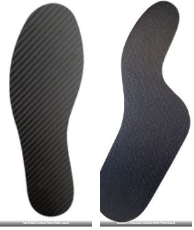
Painful conditions of the forefoot can often benefit from rigid carbon plate insoles that make the shoe stiffer to restrict motion and decrease pain. This is particularly helpful for osteoarthritis or hallux rigidus of the hallux joint, turf toe and Freiberg’s infarction.

There is two different types of these rigid carbon plates based on their width in the forefoot. The full width one is rigid across the entire forefoot where as the one with what is called a Morton’s extension only goes under the big tie which allows the other side of the forefoot to have some more movement.

These inserts are only 1.0 to 1.2mm thick so easily fit into the shoes without any issues. They do change the way you walk, so may take a while for people to get used to wearing them.